Discover how blood is carried away from the heart, and learn which blood vessels carry blood to and from the heart. Understand the difference between arteries, veins, and vessels.
Updated: 11/21/2023
Major Blood Vessels That Carry Blood Away From the Heart
Table of Contents
Show
Frequently Asked Questions
What are the two main blood vessels that carry blood to the heart?
The two main blood vessels that carry blood to the heart are the pulmonary vein, which carries oxygenated blood from the lungs back to the heart, and the vena cava, which carries deoxygenated blood from the tissues back to the heart.
Which blood vessels carry blood to and from the lungs?
The pulmonary artery carries deoxygenated blood to the lungs, and the pulmonary vein carries oxygenated blood back to the heart.
Which are the two arteries that carry blood away from the heart?
The pulmonary artery carries deoxygenated blood to the lungs, and once it returns to the heart (via the pulmonary vein), oxygenated blood is pumped out to the rest of the body through the largest artery in the body, the aorta.
What blood vessels carry blood away the heart?
The blood vessels that carry blood away from the heart are known as arteries. Arteries usually carry oxygen-rich blood, but one artery, the pulmonary artery, carries deoxygenated blood from the heart to the lungs.
Table of Contents
ShowOn average, it takes about a minute for blood to leave the heart, circulate throughout the body, and return to the heart to be pumped back out again. What carries all that blood throughout the entire body? There is a complex network of blood vessels that takes blood from the heart to the rest of the body. The blood vessels that carry blood away from the heart are known as arteries, while those that carry blood back to the heart are veins.
When blood first returns to the heart, it is very low in oxygen, so the first journey it makes is from the heart to the lungs, where it can pick up oxygen and get rid of carbon dioxide. The pulmonary artery is a large artery that carries this deoxygenated blood to the lungs. After passing through tiny capillaries in the lungs and becoming oxygenated, blood travels back to the left atrium of the heart via the pulmonary veins. The pulmonary arteries and veins are different from the rest of the circulatory system because the pulmonary artery is the only artery to carry deoxygenated blood, and the pulmonary vein is the only vein to carry blood that contains a lot of oxygen.
After returning from the lungs, freshly oxygenated blood enters the left atrium of the heart. From there, it flows into the left ventricle, and when the left ventricle contracts with each heartbeat, blood is pushed out of the heart and into the largest artery in the body, the aorta. The left ventricle is much thicker than the right ventricle because it has to generate much higher pressures in order to pump blood throughout the entire body.
The aorta is a very large artery with thick, muscular walls. It must be able to withstand high pressures and allow a tremendous amount of blood to pass through it without leaking. The aorta branches into several smaller arteries, and these keep branching and getting smaller and smaller until they reach capillaries, which are so tiny that they are only about the width of a single blood cell. This is where oxygen and carbon dioxide exchange can occur. Once blood leaves capillaries, it enters into the other side of the circulatory system, which is made up of veins that return the blood back to the heart.
 |
Elastic Arteries
Elastic arteries are the large arteries that are directly connected to the heart. There are only two elastic arteries including the pulmonary artery and the aorta. What makes these arteries different from other arteries is that they contain a large amount of elastic tissue, which allows them to maintain a relatively constant pressure. This is important because, without this elastic tissue, the pressure in these arteries would be seriously affected by the beating of the heart. Elastic arteries have very thick, strong walls to maintain constant pressure without expanding and contracting very much.
 |
When blood first leaves the left ventricle of the heart, it enters the ascending aorta. In this region, the coronary arteries that supply the heart with blood branch off. These arteries are very important, but they are also quite small, so they can get blocked with plaque over time, leading to chest pain or a heart attack. At the top of the ascending aorta is the aortic arch. Blood vessels that supply blood to the upper body, including the arms, head, and neck, branch off of the aortic arch. The aorta then bends down to form the descending aorta, which supplies blood to the lower body.
Because the aorta is an elastic artery, its walls contain a lot of elastic tissue. This allows it to stretch, but it's important that the amount of stretch in the aorta is carefully monitored because, if it stretches too much, the walls can become damaged. Inside the walls of the aorta, there are specialized receptors called baroreceptors that sense how much the walls are stretching. This information is then used to help regulate and maintain appropriate blood pressure.
Muscular Arteries
Once blood leaves the aorta, it enters muscular arteries, which are still relatively large blood vessels that deliver blood to specific areas of the body. For example, the femoral artery delivers blood to the legs and the carotid artery delivers blood to the brain. Within the wall of muscular arteries, there is a thick layer of smooth muscle tissue, as well as some elastic tissue. The smooth muscle tissue in muscular arteries allows them to expand or contract to control the pressure in the arteries or direct more blood to certain areas of the body.
Arterioles
Arterioles are the small arteries that branch off of muscular arteries and carry blood deeper into the tissues. Arterioles are smaller than muscular arteries and have very little elastic tissue in their vessel walls. Instead, the wall of an arteriole is composed almost entirely of smooth muscle. Arterioles travel deep into tissues and connect to the tiny capillaries where gases and nutrients are exchanged.
Your next lesson will play in
10 seconds
Once blood leaves the arterioles and travels through the capillaries, it gives up most of its oxygen and picks up carbon dioxide. Then, this deoxygenated blood enters the venous system, which returns blood back to the heart. It first enters tiny venules that join together into larger and larger veins, eventually delivering blood into the largest vein in the body, the vena cava, which carries deoxygenated blood back to the heart to begin the cycle all over again.
Deep Veins
Most veins are deep within the body where they cannot be easily seen through the skin. These are known as deep veins. The main difference between arteries and deep veins is that veins have thinner vessel walls with much less smooth muscle and elastic tissue. The pressure in veins is generally much lower than that in arteries, so they don't need to have such rigid walls. As a consequence, veins can be much bigger than arteries. Most of the time, approximately 75% of the total blood volume is in the veins at any given time, with only about 10-20% in the arterial system. Unlike arteries, veins also contain valves that open and close to prevent blood from flowing back away from the heart and keep it moving in the right direction.
Superficial Veins
Superficial veins are those veins that can be seen and felt just under the skin. They have the same structure as deep veins, but can sometimes swell more because they are not surrounded by muscle tissue, which helps maintain pressure on deeper veins and keeps blood moving in the right direction.
Pulmonary Veins
The pulmonary veins are a special type of vein that carries blood back to the heart after it passes through the lungs. Unlike all the other veins in the body, pulmonary veins carry oxygenated blood.
Arteries carry blood away from the heart, and veins bring blood back to the heart. The pulmonary artery carries deoxygenated blood from the right ventricle of the heart to the lungs, and the pulmonary veins carry oxygenated blood back to the heart. Then, the left ventricle pumps blood into the aorta, which is the largest artery in the body. Along with the pulmonary artery, the aorta is an elastic artery that contains a large amount of elastic tissue in its walls. This elastic tissue allows the aorta to stretch, and there are also baroreceptors that detect how much the aorta is stretching. This information is used by the nervous system to control blood pressure. Once blood leaves the aorta, it enters muscular arteries, which are relatively large arteries that contain some elastic tissue, as well as a large amount of smooth muscle tissue. Then, blood enters the smaller arterioles that transmit blood into capillaries.
Inside capillaries, gas exchange occurs, and deoxygenated blood then enters venules on the other side. These venules come together to form deep veins and superficial veins, which both have similar structures. These veins have thinner walls than arteries and also contain valves to prevent blood from flowing back away from the heart. Finally, veins carry blood into the vena cava, which is the largest vein in the body, and from there, it goes back into the right atrium of the heart so the process can start all over again!
Video Transcript
Major Blood Vessels
Like a system of roads, your circulatory system has its own large highways and smaller back roads. In this lesson, you will learn about the largest highway, or artery, in your body. Where this artery leaves your heart, it is, in fact, very large; it's about the size of a garden hose. It first rises upward from the left ventricle of your heart before making a U-turn and plunging downward, running deep inside your body and following close to your spinal column. Early in this large vessel's journey, it sends off branches that feed important organs and structures such as your heart, brain, head, neck and arms.
Before we formally introduce this major blood vessel leaving the heart, let's visit Mr. Smith's very busy widget factory. At the factory loading docks, we see Mr. Smith's truck drivers have loaded their trucks and are ready to deliver brand-new widgets to all of the stores around the country.
Aorta
 |
To do this in an efficient way, his drivers head out on the largest highway: Highway A. Your circulatory system also has a major highway, and it is called your aorta. This is the largest artery and carries blood from the heart to the body. Your body has miles and miles of arteries, and, as you can imagine, blood must come out of the heart under very high pressure. Blood is forced into the aorta from the left ventricle of your heart, and, because so much force is needed to propel blood all the way around your body, we notice that the left ventricle has a more muscular wall than the right ventricle, which has the comparatively smaller job of pumping blood the short distance to the nearby lungs. We already learned that the aorta is about the size of a garden hose, and this large size helps distribute some of the pressure. The aorta also contains many elastic fibers that allow it to expand when blood first enters and then relax as the blood moves along. This ability to stretch and relax makes the flow of blood smoother as it travels through your circulatory system.
Your aorta is one long, continuous tube with many branches that come off of the vessel like back roads would come off of a highway. As it courses through your body, your aorta is assigned different names along its path. The ascending aorta is the first section of the aorta and contains blood from the left ventricle of the heart.
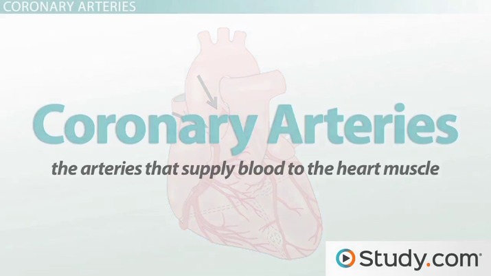
Coronary Arteries
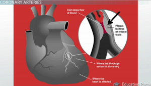 |
We don't have to travel far along the ascending aorta before reaching the first branching roads, and these are called your right and left coronary arteries. The coronary arteries are the arteries that supply blood to the heart muscle. You can imagine that the heart muscle is one of the most important muscles in your body, and therefore, your circulatory system wastes no time supplying it with richly oxygenated blood straight off of the aorta. An interesting fact about the coronary arteries is that they are relatively narrow, and these are the arteries that can become hardened and clogged due to buildup of plaque on their inner walls. If the blood flow through the coronary arteries is blocked, it can cause pain, and we call that angina. Or, if they're blocked more completely, it can result in a heart attack.
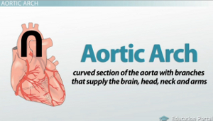
Aortic Arch
As we continue to follow the aorta away from the heart, we see that it resembles an upside-down 'U.' This section is called the aortic arch, and it's the curved section of the aorta with branches that supply the brain, head, neck and arms. A good way to think of the aortic arch is to think of it as the origin for most of the blood supply that supplies everything from your arms up to the top of your head.
 |
Baroreceptors
As we mentioned earlier, the walls of the aorta contain elastic fibers that allow it to stretch and relax along with the movement of blood through the vessel. The amount of stretching is monitored by special receptors called baroreceptors. These baroreceptors are special receptors that detect how much the aorta is stretching. If the aorta stretches a lot, that means there's a lot of pressure inside of the aorta, and this message is sent to your nervous system in response to this stretch. Your body then uses this information to help to regulate your blood pressure.
Lesson Summary
Let's review: your aorta exits your heart from the left ventricle. It is the largest artery and carries blood from the heart to the body. Because blood is propelled out of your heart under high pressure, the aorta contains elastic fibers that allow it to expand and relax. The first section of the aorta is referred to as the ascending aorta. It is off of this section that we see the first branches, which are called your right and left coronary arteries. These are the arteries that supply blood to the heart muscle, and it is these arteries that can become blocked with plaque and lead to a heart attack.
As we continue to move away from the heart, we encounter the next section, called the aortic arch. This is a curved section of the aorta with branches that supply the brain, head, neck and arms. The aortic arch also contains special receptors that detect how much the aorta is stretching. These receptors are called baroreceptors. They send a signal about the stretching to your nervous system, and this helps to regulate your blood pressure.
Learning Outcomes
Following this lesson, you could be able to:
- Evaluate the structure and function of the aorta, ascending aorta, coronary arteries and aortic arch
- Spell out the causes angina and heart attacks
- Remember the definition of baroreceptors as well as the location
Register to view this lesson
Are you a student or a teacher?
Unlock Your Education
See for yourself why 30 million people use Study.com
Become a Study.com member and start learning now.
Become a MemberAlready a member? Log In
BackResources created by teachers for teachers
Over 30,000 video lessons
& teaching resources‐all
in one place.


I would definitely recommend Study.com to my colleagues. It’s like a teacher waved a magic wand and did the work for me. I feel like it’s a lifeline.
Jennifer B.
Teacher

Back

Major Blood Vessels That Carry Blood Away From the Heart Related Study Materials
Explore our library of over 88,000 lessons
Browse by subject